Cross sectional analysis plays an important role in thin film characterization. Various techniques are available at the nanoFAB for compositional and morphological analysis of mutli-layer thin film stacks, including XPS Depth-Profiling, SEM/EDX, TEM/EDX. These techniques work complementarily to each other. It is important to choose the most suitable one(s) for your characterization needs. Here, we demonstrate the required sample preparation and typical results for these different techniques from a case study of YSZ/Ti film stacks analysis.
Sample
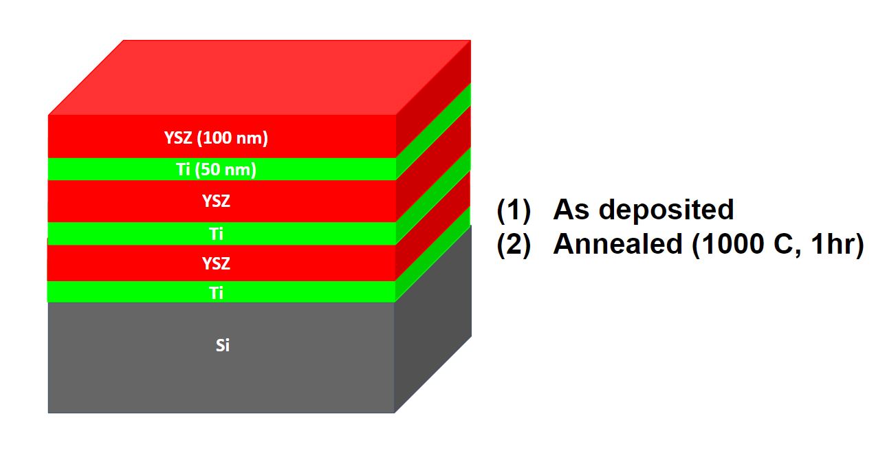
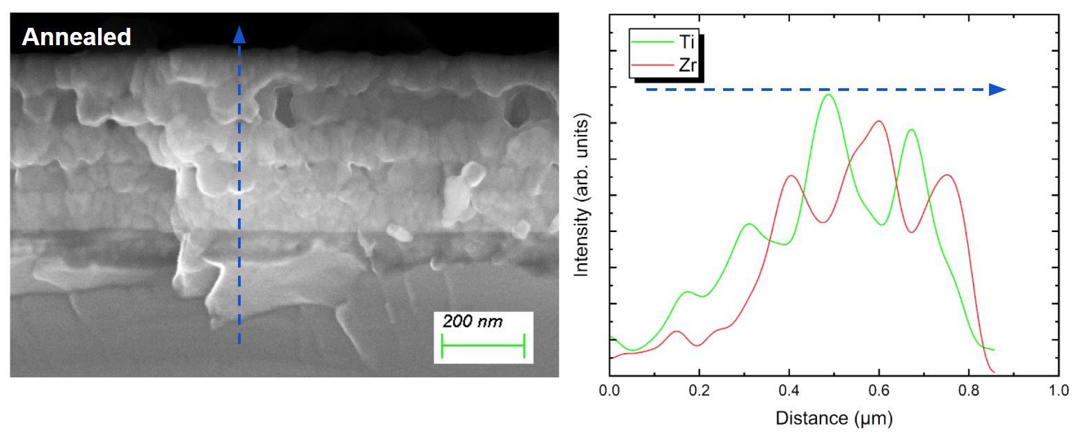
Film Stack: YSZ (100 nm) / Ti (50 nm) film stacks on Si substrates: (1) as deposited and (2) Annealed at 1000 C for 1 hour.
Sample courtesy: Avra Bandyopadhyay and Prof. Zubin Jacob, School of Electrical and Computer Engineering, Purdue University
SEM/EDX analysis of cleaved cross-section
- Sample preparation effort: ★☆☆☆☆
- Result resolution: ★☆☆☆☆
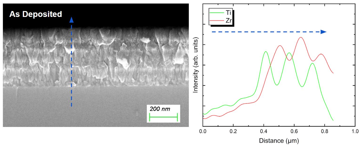
Manual cleaving is one of the simplest methods to produce cross-sections for quick SEM/EDX inspection. However, due to the unsmooth surfaces, it would be very difficult to get high resolution SEM images by Secondary Electron (SE) signals. Meaningful EDX line-profiles can be obtained, but it is very challenging to get maps.
Equipment:
Sample preparation: none
Analysis: Zeiss Sigma FESEM with Oxford EDX
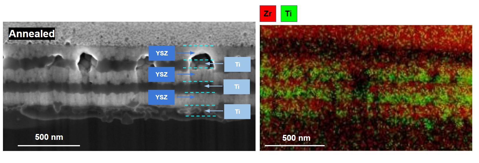
SEM/EDX analysis of ion milled cross-section
- Sample preparation effort: ★★★☆☆
- Result resolution: ★★★☆☆
Ion beam milling would produce smooth cross-sections, which enables high resolution SEM images by SE signals. Meaningful EDX maps are achievable as well.
Equipment:
Sample preparation: Thermo Fisher Helios Hydra PFIB/SEM
Analysis: Zeiss Sigma FESEM with Oxford EDX

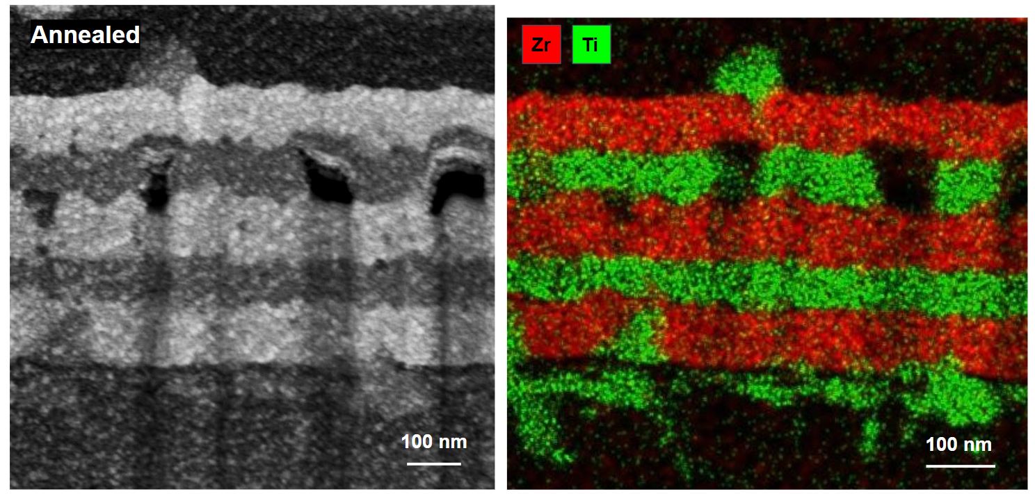
TEM/EDX analysis of thin lamella
- Sample preparation effort: ★★★★★
- Result resolution: ★★★★★
TEM analysis of thin sections/lamellas would produce the best results in terms of resolution. However, sample preparation would require a broad ion beam milling with conventional TEM sample preparation process or an FIB/SEM dual beam system that is equipped with pluckout attachments.
Equipment:
Sample preparation: Thermo Fisher Helios Hydra PFIB/SEM
Analysis: JEOL JEM-ARM200CF S/TEM with JEOL JED EDX


XPS Depth-Profiling
- Sample preparation effort: ☆☆☆☆☆
- Result resolution (lateral): ★☆☆☆☆
- Result resolution (Z): ★★★☆☆
As a complementary technique to SEM/TEM/EDX, XPS Depth-Profiling provides quantitative compositional and chemical bonding analysis with very good depth/Z resolution. Although SEM/TEM/EDX has much higher spatial resolution, EDX can only provide elemental composition information. No sample preparation is needed at all for XPS analysis. The XPS Depth-Profiling analysis is a destructive process as it utilizes ion beams to sputter materials away. Typical sputtered area is in the order of a few mm’s. The minimum analysis area (lateral resolution) is around 10 μm.
Equipment:
Sample Preparation: none
Analysis: PHI VersaProbe III Scanning Probe XPS system


In summary, SEM/EDX of manually cleaved cross sections is suitable for sample screening with simple and quick sample preparation, while SEM/EDX and TEM/EDX with ion milled cross-sections/lamellas provide the best results in terms of achievable resolution. XPS Depth-Profiling provides complementary compositional analysis.
All instruments used in this case study are open to users by either self-service or staff analysis. If you have any questions, please contact Peng Li (Peng.Li@ualberta.ca), the Characterization Group Manager. If you are interested in getting trained on any of the tools or submitting samples for staff analysis, please submit a request (training or sample) on LMACS with details of your samples and required analysis.