
The nanoFAB is pleased to announce that the Thermo Fisher Scientific Helios Hydra Plasma Focused Ion Beam / Scanning Electron Microscopy Dual Beam system has been successfully installed and is operational now.
The Helios Hydra PFIB/SEM system features:
- Multiple Ion Species Plasma Ion Source: Xe, Ar, N and O
- High resolution FESEM with sub-nm spatial resolution
- Various Imaging and Analytical detectors: SE, BSE, STEM and EDX
- MultiChem Gas Injection System (GIS): C, Pt and W
- Micro-Manipulator: precise movement and rotation
All the above features enable new characterization capabilities that were not available before:
- TEM sample preparation
- Slice & View FIB/SEM tomography
- Advanced Ion Milling with high resolution and throughput
While our team continue to commission advanced milling techniques, TEM sample preparation and standard Ion Milling are available to users as staff analysis. If you have needs for these techniques, please submit a “sample” request with sample details on LMACS. If you have any questions, please feel free to contact Peng Li (Peng.Li@ualberta.ca) – the Characterization Group Manager.
Application Examples
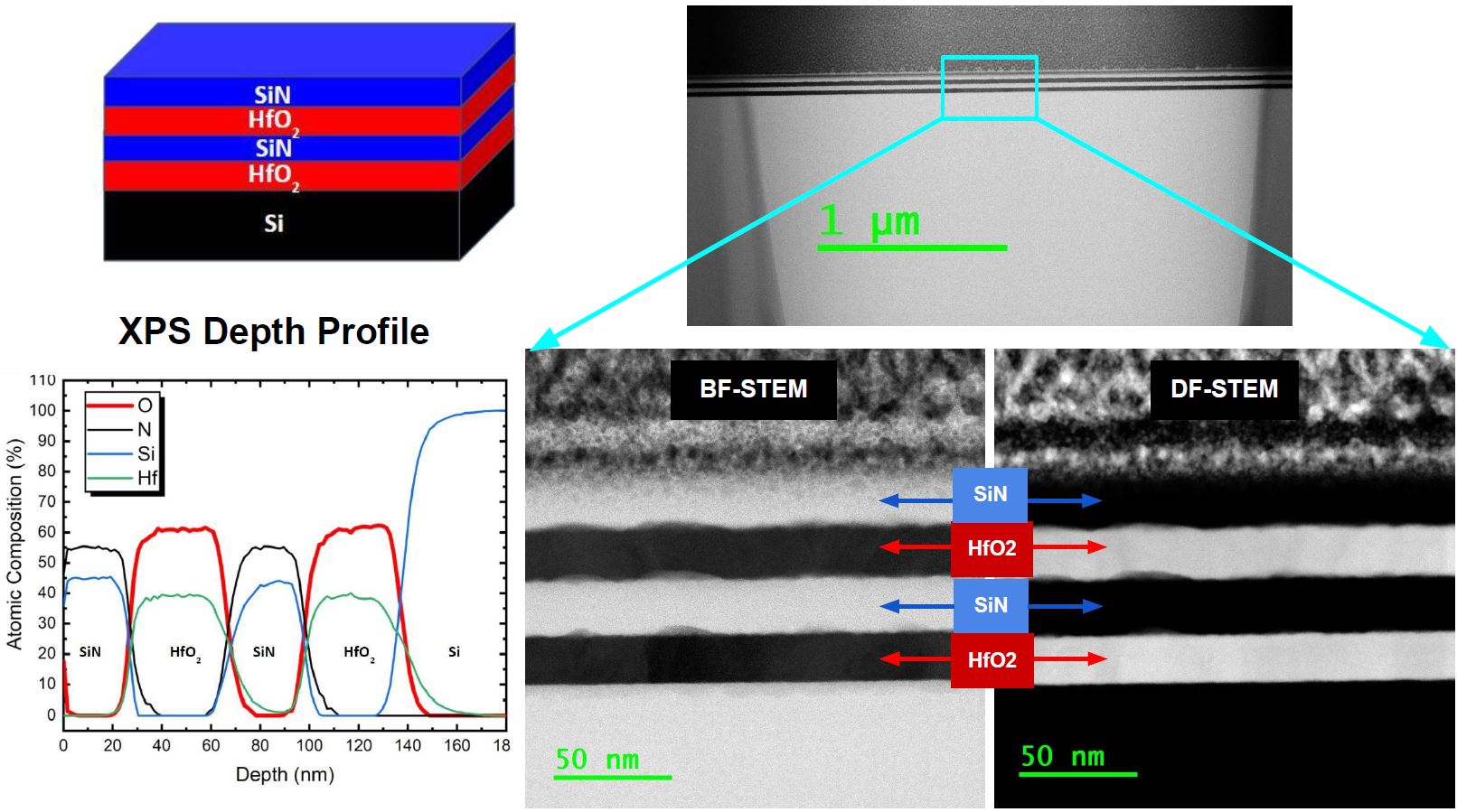
Multi-Layer Film Stack
- Sample: SiN/HfO2/SiN/HfO2 (20nm thick) films on Si
- Techniques: TEM, XPS
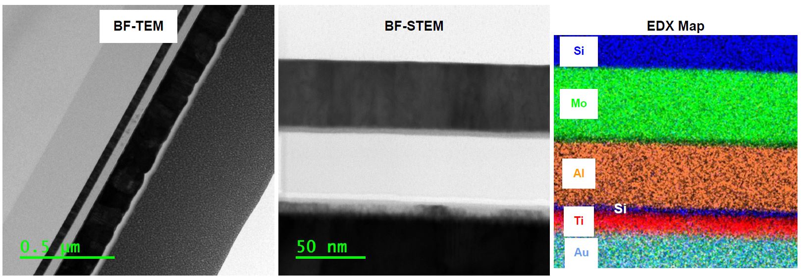
Multi-Layer Device
- Sample: Au/Ti/SiO2/Al2O3/Mo/SiO2 on Si
- Techniques: S/TEM and EDX
Cross-Section and Plan-View Analysis of NanoTubes
- Sample: TiO2 NTs on Si
- Techniques: S/TEM
- Sample Courtesy: Sheng Zeng and Prof. Karthik Shankar, Department of Electrical & Computer Engineering, University of Alberta

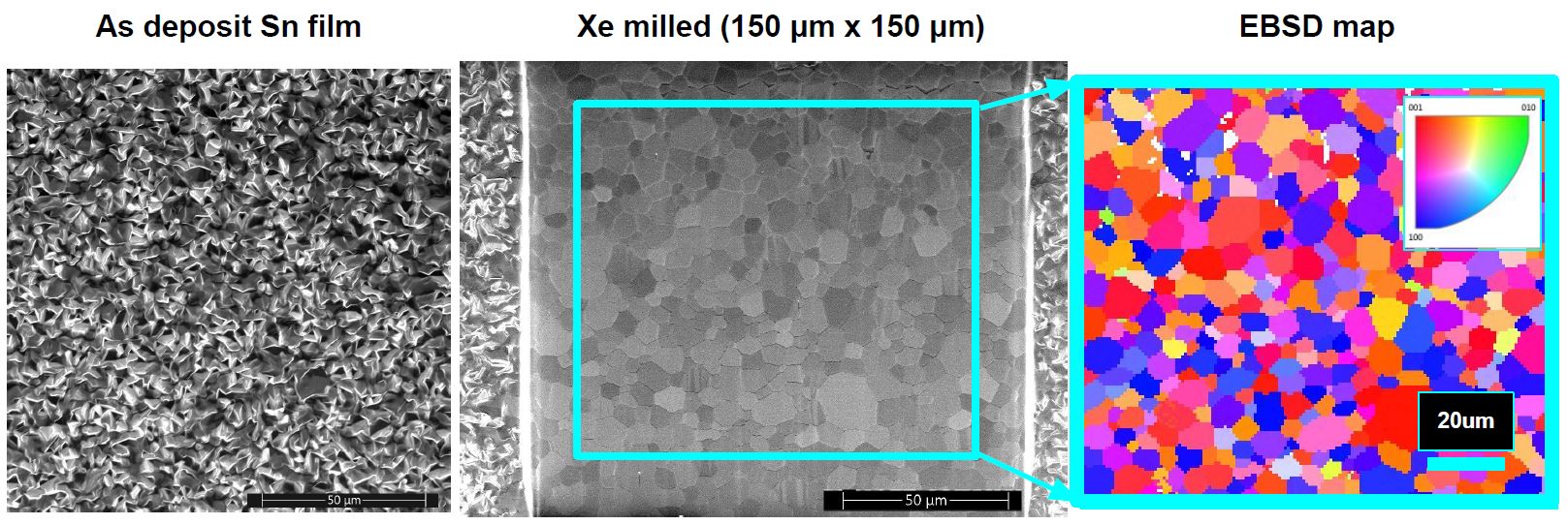
Large Area Milling for EBSD Analysis
- Sample: Sn film on Si
- Techniques: Xe milling and EBSD
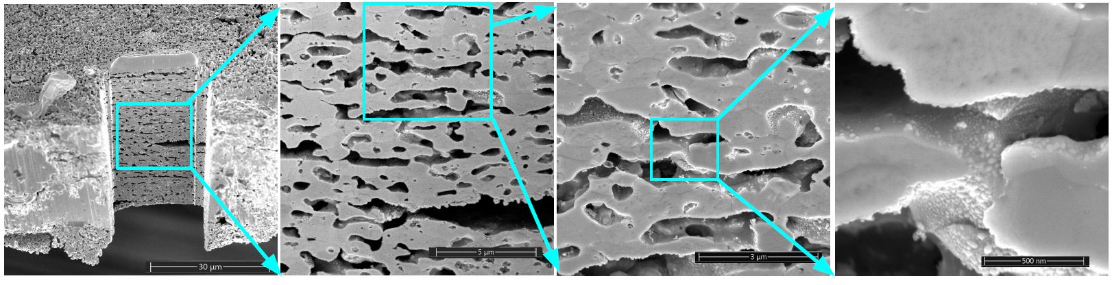
Large Area Milling for Porosity Characterization
- Sample: Porous Sn film
- Techniques: FIB and SEM
- Sample Courtesy: Dr. Peter Kalisvaart and Prof. Jillian Buriak, Department of Chemistry, University of Alberta.