Although advanced microscopy techniques are achieving very high spatial resolution (i.e. sub-nm), there has been great demand for fast and reliable nanoscopic analysis of large-area samples. Several characterization tools at the nanoFAB, which are equipped with large-area imaging/mapping capacities for both morphological and compositional analysis, have been successfully commissioned and are ready for general user work.
- Large Area High Magnification Imaging on the Helium Ion Microscope (HiM)
The NPVE system equipped on the tool has a 16 bit scan generator, and can acquire images up to 32 K x 32 K pixels. This enables acquisition of single large area images while maintaining sub-nm resolution of HiM. Below is a 20 µm x 20 µm HiM image of a Block Copolymer sample with a scan size of 16K by 16K pixels (1.2 nm/pixel). The two inset squares cover regions of 1.2 µm x 1.2 µm. (Image Courtesy: Dr. Cong Jin, Prof. Jillian Buriak Group, Department of Chemistry, University of Alberta). For details of the work, please see the paper titled ” Preferential Alignment of Incommensurate Block Copolymer Dot Arrays Forming Moiré Superstructures”, ACS Nano, 2017, 11, 3237-3246.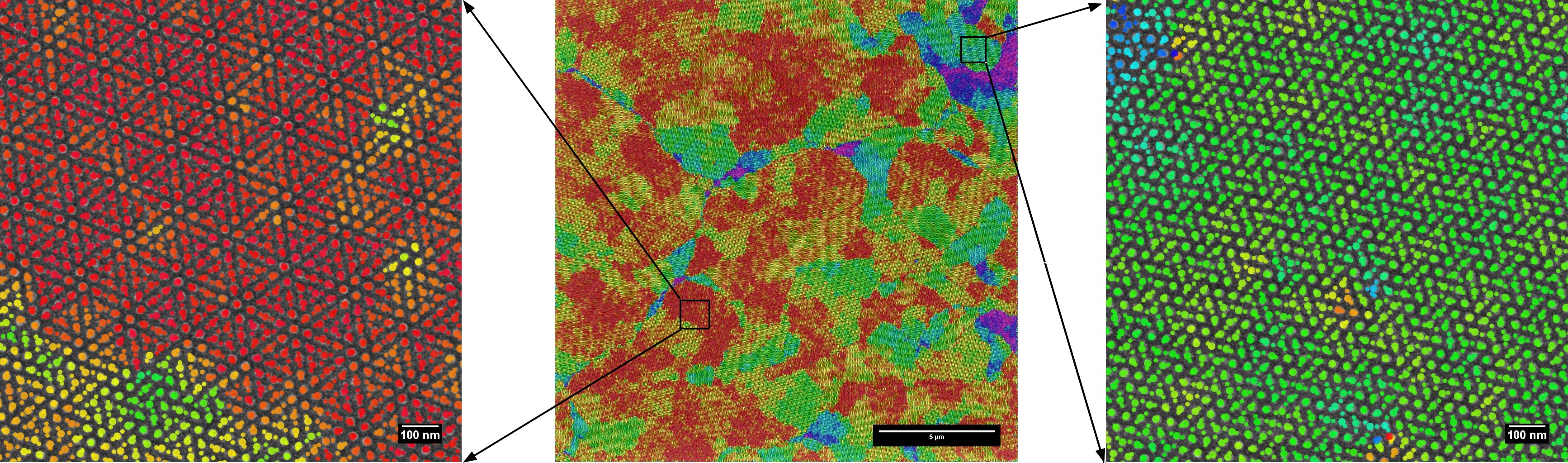
20 µm x 20 µm HiM image of a Block Copolymer sample with scan size of 16K by 16K pixel (1.2 nm / pixel)
- Large Area Elemental Mapping by X-Ray Fluorescence (XRF)
The Orbis PC Micro-EDXRF Elemental Analyzer provides easy and unattended large area chemical mapping. Maps are scanned by moving the stage. With available spot sizes (from 2 mm down to 30 µm), maps up to 124 mm x 124 mm can be completed within reasonable acquisition time. Below is a 2 cm x 1.5 cm XRF elemental map of a Geological thin section, acquired in a few hours with a 30 µm spot size. (Image Courtesy: Tyler Warchola, Earth & Atmospheric Sciences, University of Alberta).
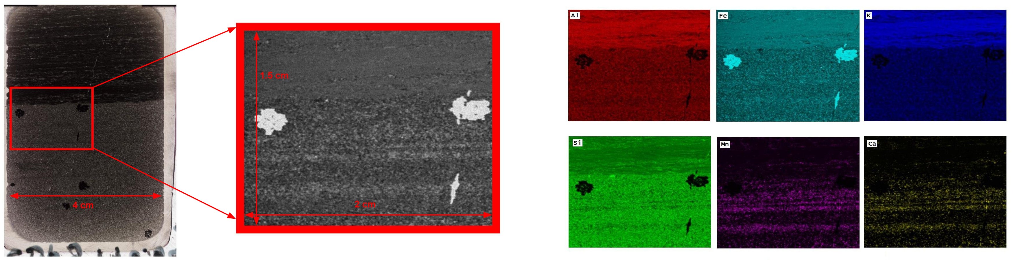
2 cm x 1.5 cm XRF elemental map of a Geological Thin Section sample.
Please contact Peng Li (Peng.Li@ualberta.ca) – the Characterization Group Manager, if you have any questions regarding the above techniques.