The nanoFAB is pleased to announce successful configuration and implementation of software solutions in our electron and ion microscopy, enabling large area imaging and correlative workflow.
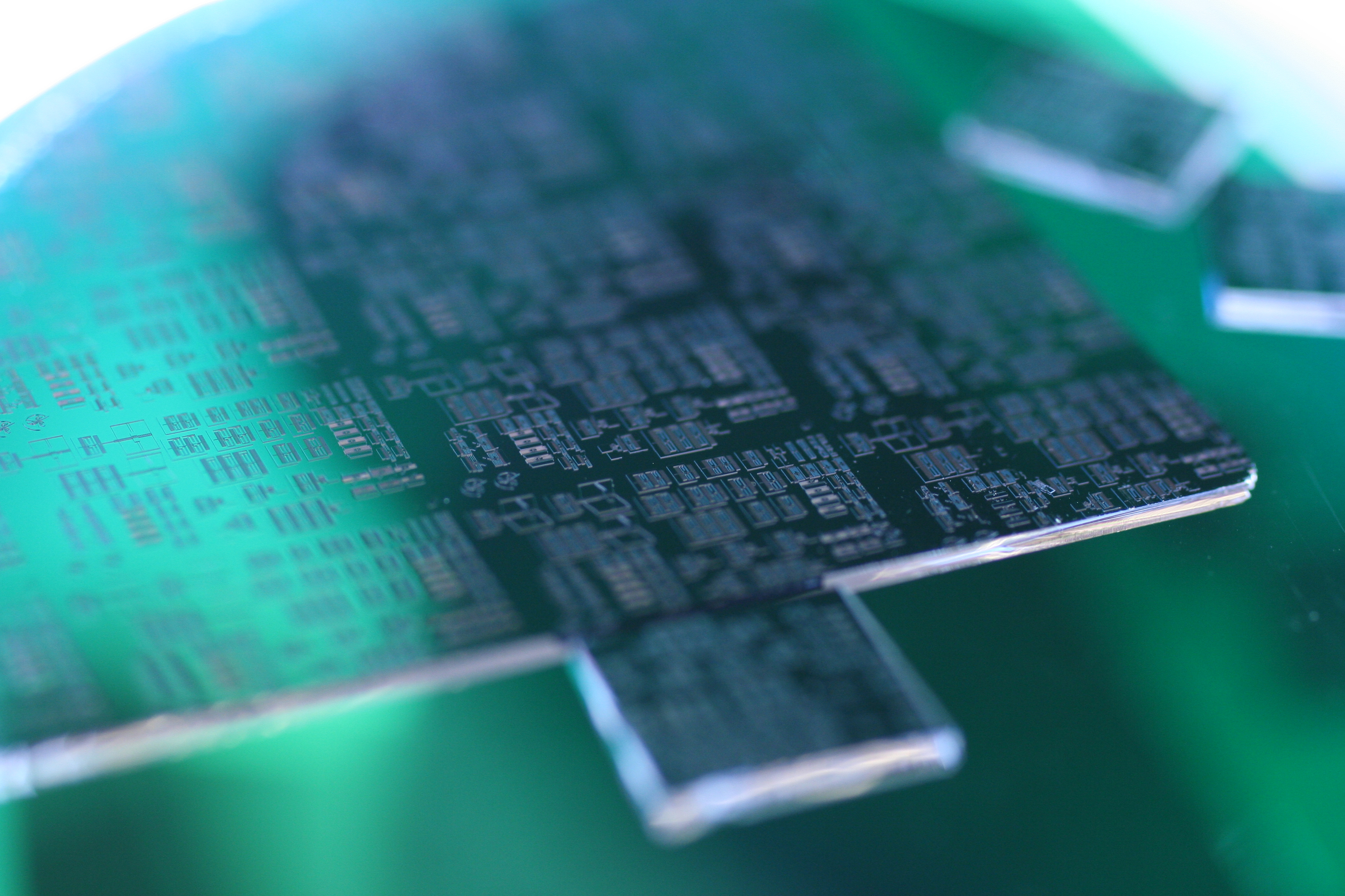
Electron & Ion Microscopy (SEM, FIB and TEM) provides morphological and compositional analysis with ultra high spatial resolution but lack of larger macroscopic context. It is also challenging to obtain analysis and observations with multiple sources from identical locations of the same devices/samples, in order to obtain comprehensive data.
Software solutions are now installed and commissioned in the following microscopes, providing large area imaging and correlative workflow:
- ATLAS on Zeiss Sigma FESEM
- ATLAS on Zeiss EVO SEM
- MAPS on ThermoFisher Hydra Plasma FIB/SEM
These software packages enable automatic workflow of multi-scale (from cm to nm), multi-platform (optical, x-ray, electron and ion microscopy and spectroscopy) and multi-dimensional (2D, 3D and 4D) characterization. Please see the application examples below of how the workflow can provide correlative analysis by linking Optical, Raman, AFM, XRM, SEM, FIB, and TEM data.
ATLAS and MAPS software are now available for user training. If you are interested in utilizing the workflow for your material characterization, please submit a training request on LMACS. If you have any questions, please feel free to contact Shihong Xu (shihongx@ualberta.ca), Josh Perkin (jperkins@ualberta.ca) or Peng Li (peng.li@ualberta.ca).
Application Examples
- Sample: Porous Ni film
- Application: Large area imaging for multi-scale analysis (Google Earth like images with high spatial resolution)
- Instrument:
- Zeiss Sigma FESEM

- Sample: Shale
- Application: Multi-scale and correlative SEM/EDX/Raman analysis of elemental and mineral distribution
- Instruments:
- Zeiss EVO SEM with Oxford EDX
- Renishaw inVia Confocal Raman

- Sample: Mineral inclusions
- Application: Multi-scale and correlative SEM/EDX/XRM/TEM analysis of mineral inclusions in magnetite-apatite deposits
This work is published in Geology (2024) 52 (6): 417–422 - Instruments:
- Zeiss Versa 620 X-Ray Microscope
- ThermoFisher Hydra Plasma FIB/SEM
- JEOL ARM S/TEM

- Sample: Multi-layer AlGaAs thin film
- Application: Correlative SEM/EDX/AFM/TEM analysis to characterize both surface and cross section morphology and composition
- Instruments:
- ThermoFisher Hydra Plasma FIB/SEM
- Bruker Dimension Edge AFM
- JEOL ARM S/TEM
