Advanced XRD techniques and applications on Bruker D8D plus diffractometer
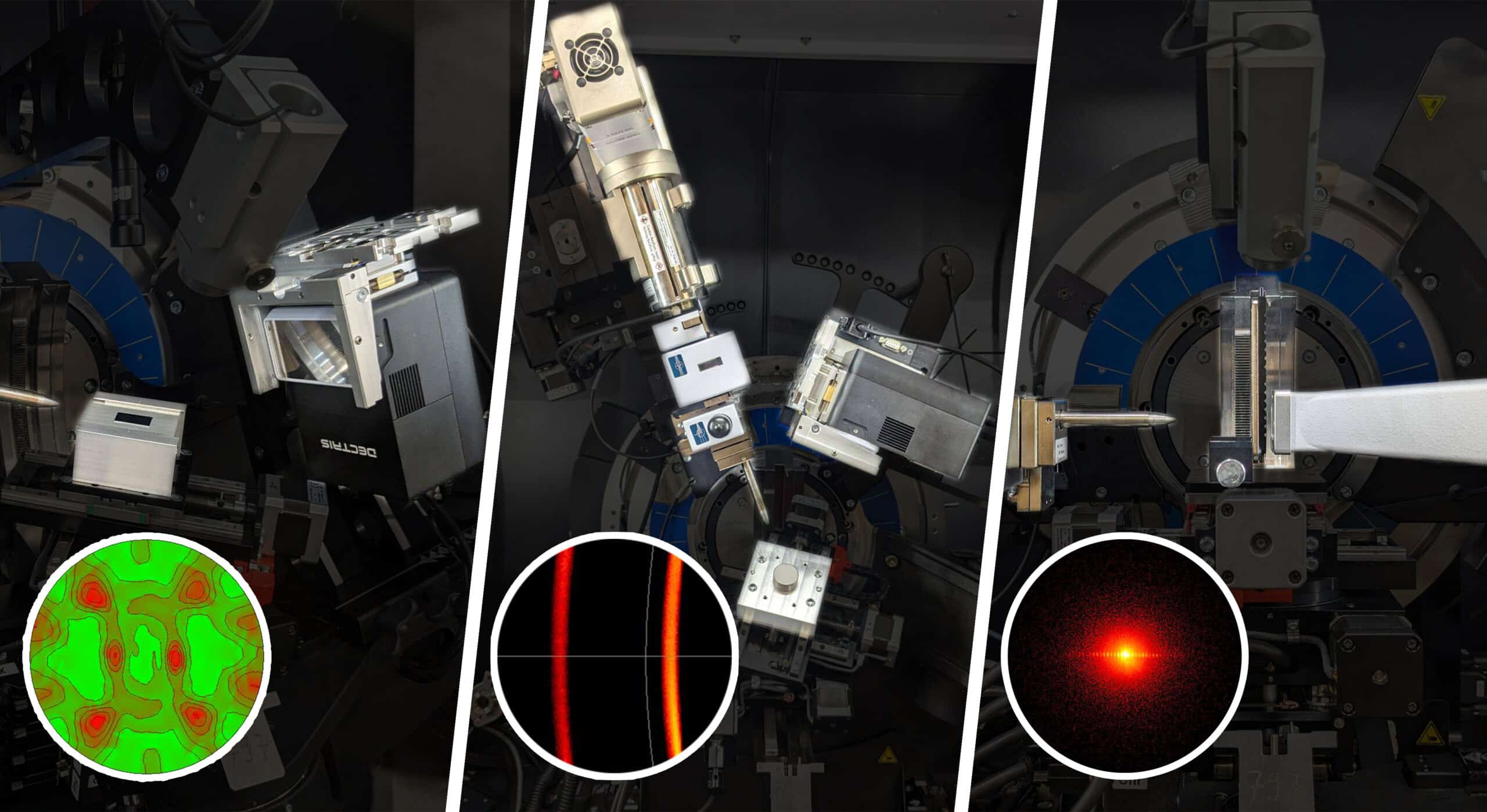
The nanoFAB is pleased to announce that advanced In-Plane Diffraction (Non-Coplanar Scan), Pole Figure Measurement, 2D Stress Measurement, and Multi-Angle Scattering (SAXS/WAXS) are fully commissioned on the Bruker D8 DISCOVER Plus X-Ray Diffractometer .
IN-PLANE DIFFRACTION (NON-COPLANAR SCAN): With standard diffraction geometries (coplanar scan), such as the Bragg-Brentano geometry, X-rays penetrate to a certain depth into the sample, and the diffraction from lattice planes parallel to the sample surface is measured. In case of ultra-thin films, X-rays completely transmit and no diffraction is observed from the thin film with the standard coplanar scan. In these circumstances, in-plane diffraction enabled by non-coplanar scan provides effective and efficient analysis, in which both the incident and diffracted beams are nearly parallel to the sample surface, as the detector scans in the plane of the horizontally positioned sample. In-plane diffraction has two major features:
(1) The penetration depth of the X-ray is limited to the top surface of the sample. By setting the X-ray incidence at the critical angle or slightly higher, ultra–thin films and the texture of surface layers can be analyzed.
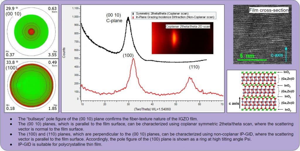
(2) The technique measures lattice planes that are (nearly) perpendicular to the sample surface, which are inaccessible by other techniques. This second feature is a key difference between the in-plane grazing incidence diffraction (IP-GID) vs. the conventional GID with coplanar scan. Figure 1 shows the setup of in-plane diffraction (non-coplanar scan) on the Bruker D8D plus XRD, as compared to coplanar symmetric 2theta/theta scan for powder diffraction on the Bruker D8 XRD and coplanar GID for thin-film applications on the Rigaku XRD.
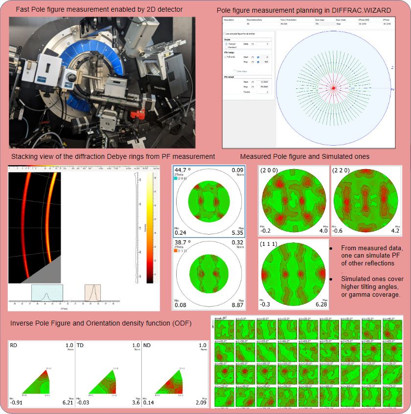
QUANTITATIVE TEXTURE ANALYSIS: The presence of crystallographic texture (preferred orientation) in polycrystalline materials has a significant effect on the anisotropy of the materials properties. Therefore, it is critically important to obtain a qualitative/quantitative description of the orientation distribution of crystallites, or the orientation distribution function (ODF) in order to characterize and predict their properties. While direct measurement of the ODF is very challenging, pole figures (PF) can be measured to reconstruct the ODF experimentally, where the diffraction angle is fixed and the diffracted intensity of a certain lattice plane is record by varying two geometrical parameters, such as the alpha angle (tilt angle of the scattering vector from the surface normal direction of a sample) and the beta angle (rotation angle of the scattering vector around the surface normal direction of a sample). The sensitive and large size 2D detector available on the Bruker D8D plus provides large coverage in Gamma and 2Theta, thus high speed and high throughput PF measurement (compared to measurement using a 0D or 1D detector), by taking 2D frames as continuous scans in PHI (the azimuthal angle) at successive values of Psi (the tilt angle).
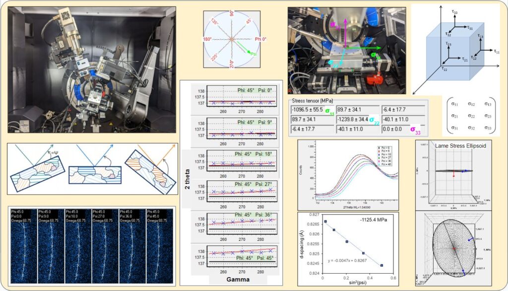
RESIDUAL STRESS ANALYSIS: Residual stress, created during the materials manufacturing process or accumulated during operation, can have serious negative effects on a product's quality, performance, and durability, as it may result in cracks and delamination of the films, as well as deformation of the substrate depending on the adhesion strength between the film and the substrate. Residual stress X-ray diffraction is one of the techniques for evaluating the near-surface residual stress with high accuracy, which is non-destructive and is applicable to polycrystalline materials with moderate to fine grain sizes. In X-ray diffraction residual stress measurement, the strain in the crystal lattice is measured using the changes in the d-spacing of the crystal lattice planes as the strain gauge, and the residual stress producing the strain is calculated assuming a linear elastic distortion of the crystal lattice. The most commonly used method for XRD stress determination is the sin2(Psi) method. By measuring the change in the d-spacing of a suitable lattice plane for at least two different tilting angles (Psi), the stress present in the plane of sample surface can be calculated from the slope of the d vs. sin2(Psi) plot. Our Bruker D8D plus XRD provides 2D stress measurement with the side inclination method, enabled by Psi tilt and PHI rotation of the Eulerian cradle, together with the large Gamma coverage of the 2D detector. 2D stress analysis measures not only the peak position but also the shape of the diffraction Debye rings. Essentially, it describes the curvature of the diffraction rings as the scattering vector tilted away from the surface normal, providing a more generalization solution than the simple sin2(Psi) peak position analysis, which is a special condition of the 2D stress analysis. The procedure of 2D stress measurement is similar to that of 2D pole figure measurement, except that a higher-angle diffraction peak needs to be used.
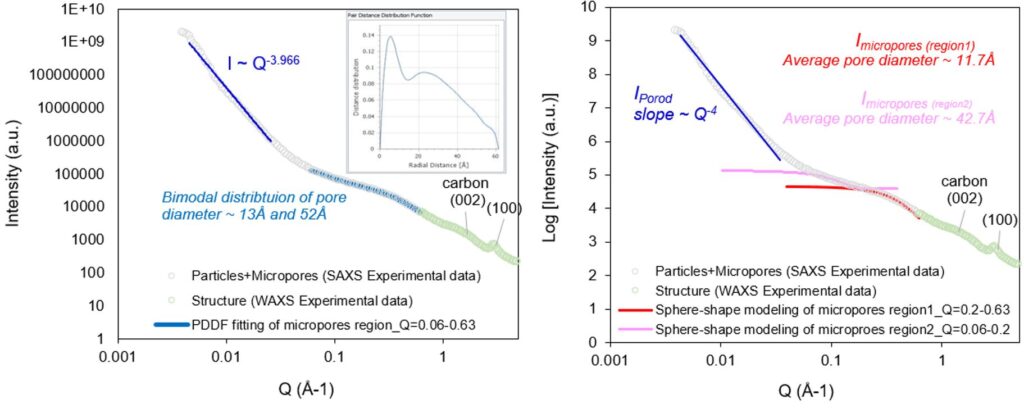
MULTI-ANGLE SCATTERING (SAXS/WAXS) ANALYSIS: Small-Angle X-ray Scattering (SAXS) analyzes the elastic scattering behavior of X-rays when traveling through the material, recording their scattering at small angles (typically 0.1 - 10°). SAXS quantifies nano-scale density differences in a sample, enabling characterization of particle sizes, shapes, distribution, pore sizes, characteristic distances of partially ordered materials, and much more. Wide-Angle X-ray Scattering (WAXS) is similar to SAXS, except that the distance from sample to the detector is shorter and thus diffraction maxima at larger angles are observed. WAXS usually covers 2theta 5-60 degree, revealing scattering/diffraction at sub-nanometer scale such as crystalline lattice planes. Multi-angle scattering analysis utilizes both SAXS and WAXS in conjunction to probe a wide range of length scale from an angstrom to a micrometer, revealing a full picture of the materials structure.
In-plane diffraction, pole figure, and 2D stress measurements are now available for user training and staff analysis. Please see the application examples and discussions below for more details. If you are interested in utilizing these techniques for your materials characterization, please submit a training request on LMACS. If you have any questions, please feel free to contact Dr. Xuehai Tan (xtan@ualberta.ca) and Peng Li (peng.Li@ualberta.ca) – the Characterization Group Manager.

Application Examples
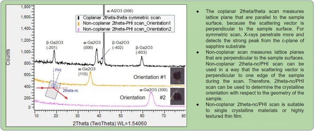
Sample: Epitaxially grown Ga2O3 thin film on sapphire substrate
Analysis: In-plane diffraction (Non-coplanar scan)
Application: Coupling non-coplanar 2theta-nc / PHI scan and coplanar 2theta/theta symmetric scan to characterize epitaxially grown Ga2O3 thin film on sapphire substrate.
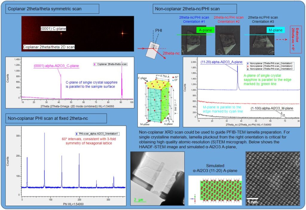
Sample: Single crystal sapphire α-Al2O3
Analysis: In-plane diffraction (Non-coplanar scan)
Application: Coupling different non-coplanar scans and coplanar symmetric scan to characterize single crystalline materials. An example is given on how in-plane diffraction could be used to guide PFIB-TEM lamella preparation.
Sample: Soda can Al sheet produced through cold-rolling
Analysis: Fast pole figure measurement enabled by 2D detector
Application: Texture analysis of the orientation distribution of crystallites from pole figure measurements.
Sample: Fiber-textured In-Ga-Zn-O (CAAC-IGZO) film on Si substrate for 3D glasses-free holographic displays
Analysis: Pole figure, coplanar symmetric scan, non-coplanar in-plane GID, PIB-STEM
Application: A correlative study of fiber-textured IGZO film using pole figure, coplanar and non-coplanar XRD (In-plane GID), and PFIB-STEM microscopy of the film cross section.
Sample Courtesy: Avalon Holographics Inc., Edmonton
Sample: Fe film with compressive stress
Analysis: 2D stress analysis
Application: A more generalization residual stress analysis solution than the simple sin2(Psi) peak position analysis
Sample: A hard carbon with rich closed pores.
Analysis: Multi-angle X-ray scattering
Application: Coupling SAXS and WAXS to investigate the internal pore sizes and the short-range ordered carbon structure.
